

Novel Wireless Sensor Monitors Progression of Soft Tissue Disorders
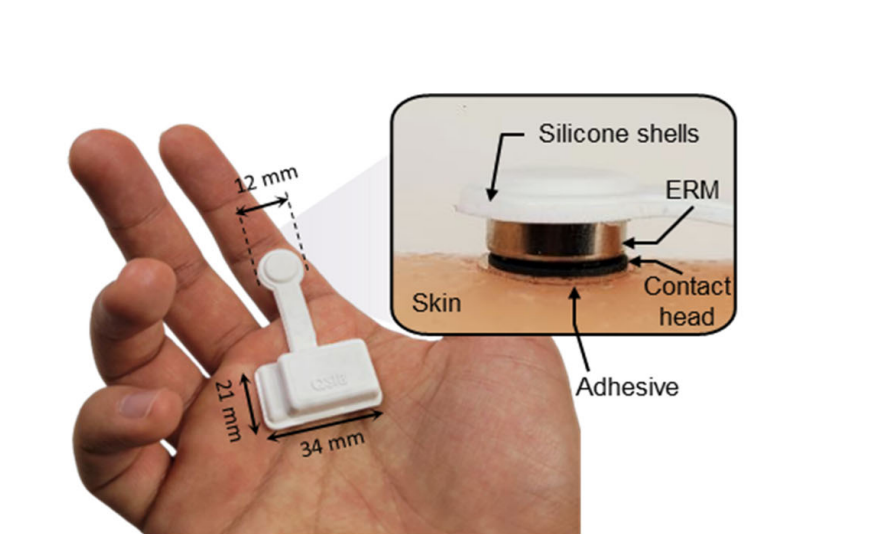
Northwestern scientists have developed a novel wireless sensor device that can continuously monitor skin elasticity in patients with soft tissue disorders including lymphedema and scleroderma, which can support early disease detection and better inform care strategies, as detailed in a study published in Science Advances.

The sensor, which is directly applied to the patient’s skin, can measure a patient’s tissue elasticity and then wirelessly transmit critical disease progression data, enabling remote access of that data by a healthcare provider from the patient’s own home and potentially omitting the need for an in-person clinical examination.
“The great thing about the sensor is it’s small, the potential for home use is tremendous, and for patients who know that they are at risk and know that they need to be constantly monitoring and detecting, this could give them a really good home-based unit that could potentially communicate data directly to their physician. That’s our goal,” said Ann Marie Flores, PT, MSPT, MA, PhD, CLT, FAPTA, associate professor of Physical Therapy and Human Movement Sciences and of Medical Social Sciences in the Division of Implementation Science, who was a co-corresponding author of the study.
Current diagnostic approaches for lymphedema — chronic tissue swelling caused by damage to lymph vessels resulting from infection, cancer treatment or, in some cases, genetic mutations — include measuring volume of the affected body part, extracellular fluid via bioimpedance spectroscopy or tissue dielectric constant and a thorough clinical exam. There is no cure for lymphedema, and continuous monitoring is essential to mitigate disease progression and prevent infection.

Skin stiffness that is caused by lymphedema is measured with an indentation test (pressing on the swollen area to measure for fluid buildup), a skin suction test or elastography imaging. While these techniques have shown to be effective, they require the use of bulky instruments, hands-on clinical examination, and are less sensitive as lymphedema progresses.
The new device, which utilizes technology developed by the laboratory of John Rogers, PhD, the Louis Simpson and Kimberly Querrey Professor of Materials Science and Engineering, Biomedical Engineering and Neurological Surgery, is flexible and can be directly applied to the patient’s skin across different parts of the body.
The device emits vibrations via an accelerometer and, from these vibrations, collects information about the skin’s stiffness, elasticity and resistance to pressure. This data can then be wirelessly transmitted from the device to the patient’s healthcare provider.
“The device can wirelessly transmit the data and then we can directly analyze this vibrating amplitude and frequency to know the skin modulus value,” said Min-Seung Jo, PhD, a postdoctoral fellow in the Rogers laboratory and lead author of the study.
The device was also validated in patients with cancer-related lymphedema and systemic scleroderma, a rare autoimmune disorder in which excess collagen production causes patches of the skin to thicken abnormally.
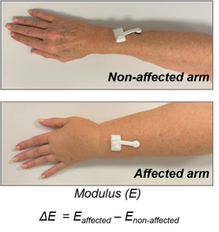
In both studies, the investigators found that the device’s larger contact area enabled deeper tissue monitoring, which improved overall evaluation of disease severity and progression. The device also demonstrated a stronger correlation compared to commercially available clinical devices that use bioimpedance and tissue dielectric constant to detect the presence of early developing lymphedema.
Unlike other methods, particularly bioimpedance spectroscopy, the device was also sensitive enough to distinguish between three different stages of lymphedema. The investigators also found the device’s larger contact area improved the measurement of skin stiffness during muscle movement and relaxation in the patients.
While diagnosis requires clinical expertise to diagnose and stage lymphedema, the new device would allow patients and healthcare providers to monitor a patient’s disease progression while the patient is in their comfort of their own home, improving early detection, accessibility and overall care, according to the authors.

“Our next steps are to see how we can make this truly home-based, how adherent patients are [with lymphedema monitoring] and to hear what providers want to know,” said Flores, who is also director of Cancer Rehabilitation for the Cancer Survivorship Institute of the Robert H. Lurie Comprehensive Cancer Center of Northwestern University.
Yonggang Huang, PhD, the Achenbach Professor of Mechanical Engineering, Civil and Environmental Engineering, and Materials Science and Engineering at the McCormick School of Engineering, was a co-corresponding author of the study.
Co-authors of the study include Carrie Richardson ‘12 MD, ‘GME, associate professor of Medicine in the Division of Rheumatology; and Jodi Johnson, PhD, research assistant professor of Pathology, of Medical Social Sciences and of Dermatology.
This work was supported by the Institute of Information and Communications Technology Planning and Evaluation (IITP) grant funded by the Korea government (MSIT) (IITP-RS-2025-02214780, Generative Haptics and Fine Response Inference for Flexible Tactile Interfaces) and the Robert H. Lurie Comprehensive Cancer Center of Northwestern University under the Translational Engineering Initiative.
Legal Disclaimer:
EIN Presswire provides this news content "as is" without warranty of any kind. We do not accept any responsibility or liability for the accuracy, content, images, videos, licenses, completeness, legality, or reliability of the information contained in this article. If you have any complaints or copyright issues related to this article, kindly contact the author above.
